Embark on a journey to unravel the complexities of the superior view of the foot, where bones, muscles, and nerves intertwine to orchestrate movement and support. From common injuries to surgical interventions, this comprehensive guide delves into the fascinating world beneath our feet.
Unveiling the intricate anatomy, we’ll explore the bones and their articulations, the muscles and their insertions, and the intricate network of nerves and blood vessels that animate the foot. We’ll delve into the clinical significance of the superior view, examining common injuries, pathologies, and the crucial role of imaging in diagnosis and treatment.
Anatomy of the Superior View of the Foot
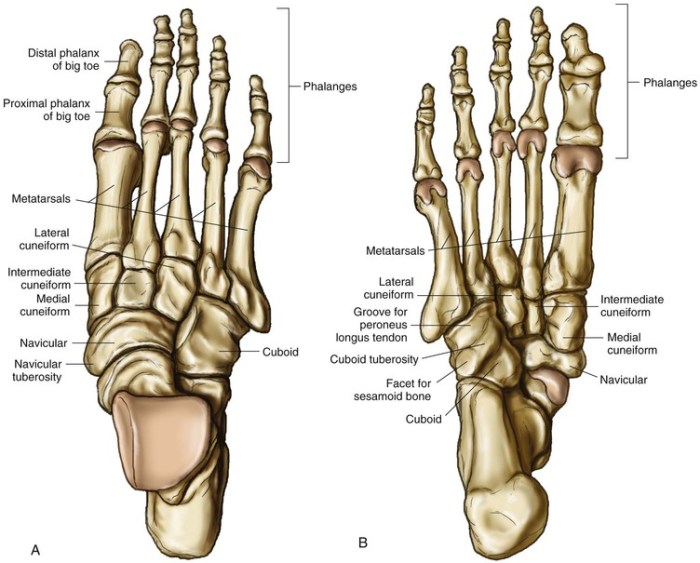
The superior view of the foot presents a complex arrangement of bones, muscles, nerves, and blood vessels. Understanding the anatomy of this region is crucial for comprehending foot mechanics and managing various foot conditions.
Bones and Articulations
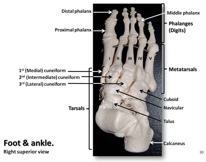
The superior view of the foot reveals the intricate arrangement of tarsal and metatarsal bones.
In the superior view of the foot, the medial and lateral sesamoids are positioned just proximal to the distal aspect of the third metacarpal bone. As with other equine breeds, the hooves of horses with a mottled coat are slightly oval in shape and the dorsal hoof wall is often steeper than in other breeds.
In the superior view of the foot, the coronary band is clearly visible.
- Tarsal Bones:The tarsal bones, comprising the talus, calcaneus, navicular, cuboid, and three cuneiforms (medial, intermediate, and lateral), form the posterior and midfoot region. These bones articulate with each other to provide stability and mobility to the foot.
- Metatarsal Bones:The five metatarsal bones extend anteriorly from the tarsal bones and form the forefoot. They articulate with the tarsal bones proximally and the phalanges distally.
Muscles and Insertions
The superior view of the foot demonstrates the presence of several muscles responsible for dorsiflexion, eversion, and inversion movements.
- Extensor Digitorum Longus:Originates from the anterior surface of the tibia and fibula; inserts onto the dorsal surface of the phalanges, extending the toes.
- Extensor Hallucis Longus:Originates from the anterior surface of the fibula; inserts onto the dorsal surface of the hallux (big toe), extending the big toe.
- Peroneus Tertius:Originates from the anterior surface of the fibula; inserts onto the base of the fifth metatarsal, everting the foot.
- Tibialis Anterior:Originates from the lateral condyle of the tibia; inserts onto the medial cuneiform and base of the first metatarsal, inverting the foot.
Innervation and Blood Supply
The superior view of the foot receives innervation from various nerves and blood supply from several arteries.
- Innervation:The superior view of the foot is innervated by branches of the deep peroneal nerve (L4-S1) and the superficial peroneal nerve (L5-S2).
- Blood Supply:The dorsalis pedis artery, a branch of the anterior tibial artery, supplies blood to the superior view of the foot.
Clinical Significance of the Superior View of the Foot
The superior view of the foot provides valuable insights into various foot pathologies and injuries. Understanding the clinical significance of this perspective aids in accurate diagnosis, appropriate treatment planning, and effective rehabilitation.
Common Injuries and Pathologies
- Plantar fasciitis:Inflammation of the plantar fascia, a thick band of tissue that supports the arch of the foot.
- Heel spurs:Bony growths that develop on the underside of the heel bone, often associated with plantar fasciitis.
- Morton’s neuroma:A benign nerve entrapment that causes pain and numbness between the toes.
- Tarsal tunnel syndrome:Compression of the posterior tibial nerve as it passes through the tarsal tunnel, resulting in pain and paresthesia.
Role of Imaging in Diagnosis
Imaging techniques, such as X-rays, MRI, and ultrasound, play a crucial role in diagnosing foot injuries and pathologies. X-rays can reveal bony abnormalities, such as heel spurs or fractures. MRI provides detailed images of soft tissues, including ligaments, tendons, and nerves, aiding in the diagnosis of conditions like plantar fasciitis and Morton’s neuroma.
Ultrasound can visualize fluid collections, tendon tears, and nerve entrapment.
Principles of Treatment and Rehabilitation
Treatment for foot injuries and pathologies varies depending on the specific condition. Conservative measures, such as rest, ice, compression, and elevation (RICE), are often the first line of treatment. Physical therapy can help improve flexibility, strength, and range of motion.
In some cases, injections of corticosteroids or other medications may be necessary. Surgery may be considered for severe or persistent injuries that do not respond to conservative treatment.
Surgical Approaches to the Superior View of the Foot
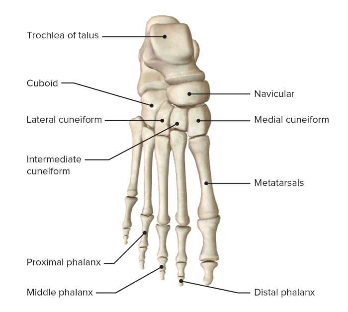
Surgical approaches to the superior view of the foot involve accessing the dorsal aspect of the foot to treat various conditions. These techniques offer surgeons a direct path to the underlying structures, allowing for precise intervention.
- Dorsal Incision:This approach involves a straight incision along the midline of the foot’s dorsum. It provides excellent exposure to the extensor tendons, dorsal vessels, and nerves.
- Lateral Incision:A lateral incision is made along the lateral aspect of the foot, providing access to the peroneal tendons, lateral malleolus, and surrounding structures.
- Medial Incision:A medial incision is made along the medial aspect of the foot, offering exposure to the tibialis anterior tendon, medial malleolus, and adjacent structures.
Indications:Surgical approaches to the superior view of the foot are indicated for a variety of conditions, including:
- Tendon injuries (e.g., extensor tendon rupture)
- Ligament tears (e.g., dorsal talonavicular ligament tear)
- Fractures (e.g., dorsal metatarsal fracture)
- Ganglion cysts
- Infections
Risks:As with any surgical procedure, there are potential risks associated with these approaches, such as:
- Infection
- Bleeding
- Nerve damage
- Scarring
Benefits:The benefits of surgical approaches to the superior view of the foot include:
- Direct access to the underlying structures
- Precise and targeted intervention
- Improved visualization and illumination
Postoperative Care:Postoperative care after surgery typically involves:
- Wound care and dressing changes
- Immobilization or restricted activity
- Pain management
- Physical therapy
Complications:Potential complications following surgery may include:
- Delayed wound healing
- Infection
- Stiffness or reduced mobility
- Persistent pain
Imaging of the Superior View of the Foot
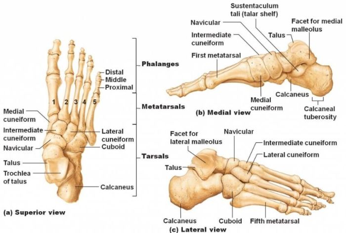
Imaging of the superior view of the foot is essential for diagnosing and managing various foot conditions. Different imaging modalities offer unique advantages and disadvantages, providing complementary information for comprehensive evaluation.
Radiography
Radiography, commonly known as X-rays, is a widely used and cost-effective imaging technique. It provides a clear visualization of bony structures, including the bones of the foot and ankle. Radiographs can detect fractures, dislocations, arthritis, and other bony abnormalities.
Computed Tomography (CT) Scan
CT scans utilize X-rays and computer processing to create cross-sectional images of the foot. They offer a more detailed view of the bones, soft tissues, and blood vessels compared to radiographs. CT scans are particularly useful for evaluating complex fractures, bone tumors, and assessing the extent of soft tissue injuries.
Magnetic Resonance Imaging (MRI), Superior view of the foot
MRI uses magnetic fields and radio waves to produce detailed images of the soft tissues of the foot. It provides excellent visualization of ligaments, tendons, muscles, and cartilage. MRI is valuable for diagnosing sprains, tears, and other soft tissue injuries, as well as detecting bone marrow abnormalities.
Ultrasound
Ultrasound uses high-frequency sound waves to create real-time images of the foot. It is commonly used to evaluate soft tissue structures, such as tendons, ligaments, and muscles. Ultrasound can also assess blood flow and detect fluid collections or foreign bodies.
Physical Examination of the Superior View of the Foot
A comprehensive physical examination of the superior view of the foot involves a thorough assessment of the foot’s structure, function, and any abnormalities. It is crucial for identifying underlying conditions and guiding appropriate treatment plans.
Steps of the Examination
The physical examination typically follows a step-by-step approach:
- Inspection:Visual examination of the foot from above, assessing its shape, size, and alignment.
- Palpation:Manual examination of the foot to assess the skin texture, temperature, and any areas of tenderness or swelling.
- Range of Motion:Evaluation of the foot’s ability to move in different directions, including dorsiflexion, plantar flexion, inversion, and eversion.
- Specific Tests and Maneuvers:Performance of specific tests and maneuvers to assess specific structures and functions, such as the Lachman test for anterior cruciate ligament (ACL) integrity.
- Neurological Examination:Assessment of sensation, reflexes, and motor function in the foot.
Interpretation of Findings
The findings of the physical examination are interpreted in conjunction with the patient’s history and other diagnostic tests. Abnormalities in the superior view of the foot may indicate underlying conditions such as:
- Deformities (e.g., bunions, hammertoes)
- Trauma (e.g., fractures, dislocations)
- Infections (e.g., cellulitis, abscesses)
- Neuropathies (e.g., diabetic neuropathy)
- Arthritis (e.g., gout, rheumatoid arthritis)
A comprehensive physical examination of the superior view of the foot is essential for accurate diagnosis and appropriate management of foot conditions.
Comparative Anatomy of the Superior View of the Foot
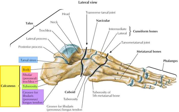
The superior view of the human foot presents a distinct arrangement of bones, joints, and muscles compared to other species. These variations reflect the unique adaptations of the human foot for bipedalism, allowing us to walk upright.
Evolutionary Significance of Anatomical Differences
The human foot has evolved over millions of years to support the weight of the body and propel us forward. The development of a longitudinal arch and a rigid heel bone (calcaneus) has provided stability and shock absorption during locomotion.
The presence of a large, opposable hallux (big toe) and a shortened lateral column have enhanced our ability to grasp and manipulate objects.
Implications for Understanding Foot Function
Understanding the comparative anatomy of the superior view of the foot is crucial for comprehending its function. The arched structure distributes weight evenly across the foot, while the rigid heel bone prevents excessive pronation. The opposable hallux assists in balance and propulsion, and the shortened lateral column allows for greater flexibility and range of motion.
Embryology of the Superior View of the Foot
The development of the foot is a complex process that begins in the embryo and continues after birth. The foot is formed from a series of mesodermal swellings that appear along the ventral surface of the embryo. These swellings eventually fuse to form the foot plate, which gives rise to the bones, muscles, and other tissues of the foot.The
molecular and genetic factors that control foot development are not fully understood, but several genes have been identified that play a role in this process. These genes include those that encode transcription factors, signaling molecules, and cell adhesion molecules. Mutations in these genes can lead to developmental abnormalities of the foot, such as clubfoot and polydactyly.Developmental
abnormalities of the foot can have a significant impact on a person’s mobility and quality of life. Clubfoot is a condition in which the foot is turned inward and downward. Polydactyly is a condition in which a person is born with extra toes or fingers.
Both of these conditions can be treated with surgery, but early diagnosis and intervention are important to ensure the best possible outcome.
FAQ Insights
What structures are visible in the superior view of the foot?
The superior view of the foot reveals the bones of the foot, including the tarsals, metatarsals, and phalanges. It also showcases the tendons and muscles that traverse the foot, along with the neurovascular structures that supply it.
What are the common injuries associated with the superior view of the foot?
Common injuries include sprains, fractures, and dislocations of the bones and joints. Tendonitis and muscle strains can also occur due to overuse or trauma.
How is imaging used in the diagnosis of superior view foot injuries?
Imaging modalities such as X-rays, MRI, and CT scans are employed to visualize the bones, soft tissues, and neurovascular structures of the foot. These techniques aid in diagnosing injuries, assessing their severity, and guiding treatment decisions.